In X-ray fluorescence spectroscopy an X-ray source is used to irradiate the sample with X-ray radiation. If the energy of this radiation is sufficient, then it will interact with the atom’s inner shell electrons causing them to be kicked out. Almost immediately, a relaxation process takes place where one of the outer shell electrons fall into the inner shell. As a result, a specific amount of energy is released in the form of electromagnetic radiation. The energy of the emitted X-rays depends on the energy difference between the higher and lower states, and therefore the radiation also carries information about the atom.

So, if we can measure the energy and the intensity of the characteristic X-ray radiation that comes out of the material during the relaxation process, the new can also get the information about the elemental composition of the sample. For measuring the energy and the intensity of the emitted characteristic X-ray radiation, there are two possible spectrometer setups. In the case of the energy-dispersive analysis, a detector is used that can sort the energies of protons. This setup is often favored due to its low cost and fast measurement times. Its main problem however is low accuracy due to broad overlapping peaks. The wavelength dispersing analysis uses a crystal to select which wavelength radiation actually enters the detector. In that crystal, the X-rays are scattered from different layers of atoms, which means that some beams travel a longer optical path. For a radiation with a defined wavelength, the criteria of Bragg’s aw is met at a certain angle and all the scattered beams are in the same place, which means that constructive interference takes place. So, if the angle is changed by moving the crystal and the detector, it is possible to scan in a wide spectral range, and find out which wavelength characteristic X-ray radiation comes out of the sample. With such a setup, the peaks are narrower and their overlapping is significantly reduced. Therefore, the accuracy in wavelength dispersive analysis is much greater than in the case of the energy dispersive analysis. The main disadvantages of this setup are longer measurement times and significantly higher cost of this spectrometer.
Now it’s time for the demonstration, where I will measure the exact elemental composition of this 300-year-old Russian coin. For that purpose, the sample is first cleaned with high-purity deionized water and organic solvents. Next, the clean substrate is placed into the spectrometer on a special holder and the experiment is started. Right now, the sample atoms are excited with X-ray radiation and the resulting characteristic X-ray’s measured. This process can take tens of minutes or even longer. Eventually, a spectrum is obtained where the energy and intensity of the emitted characteristic X-rays can be seen. Based on the data, it is possible to calculate the elemental composition of the material, and in the case of this sample we are dealing with a copper coin. The high content of oxygen and carbon originate from the rust layer, while the smaller quantities of other elements such as arsenic may indicate that the coin had a colorful history.
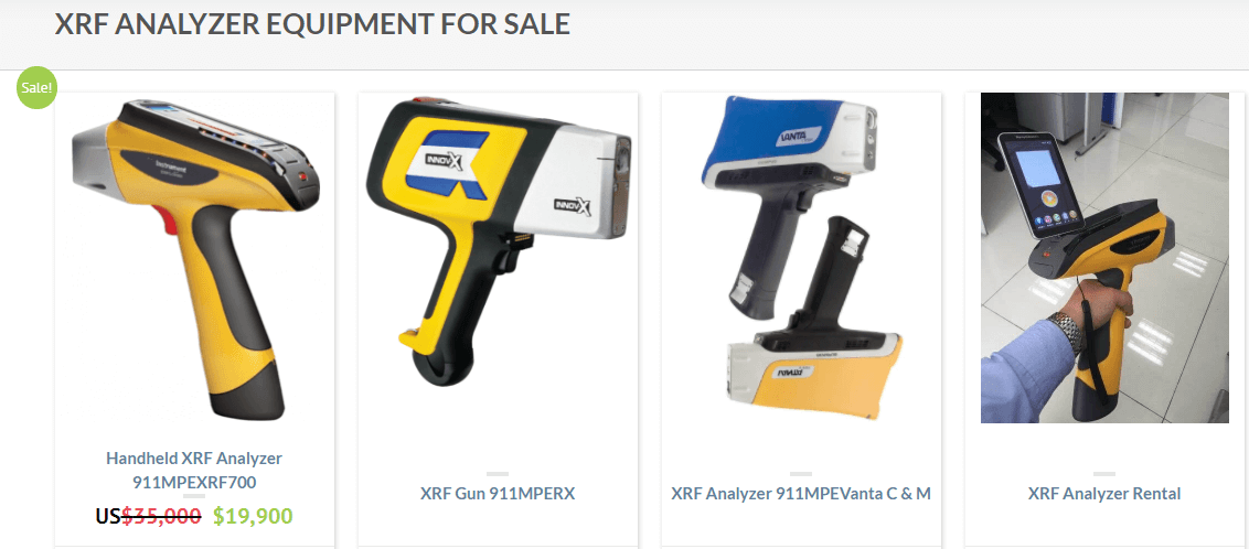
Measuring the composition of a material, however, is just one option for this system. It can also be used to map the elements on a larger substrate, measure the mass thickness of thin films or even detect very small amounts of unusual elements which can be handy in analyzing crime scene evidences. There are also smaller hand-held versions of this spectrometer in the market. Although they are less accurate than their larger cousins, they are much cheaper and more comfortable for performing quick analysis.
